Two bacterial enzymes can remove much of the blood group A signal from the blood vessels of donated human kidneys. In a study published on 30 March 2024, Serena MacMillan and colleagues tested the approach in 18 kidneys that had been declined for transplantation. The organs were studied outside the body, so the findings did not establish better survival or immediate kidney function in transplant patients.
Changing the organ’s surface
Blood groups depend on carbohydrate structures that also occur on the cells lining an organ’s blood vessels. A recipient’s antibodies can recognise incompatible structures and initiate damage to a transplanted kidney.
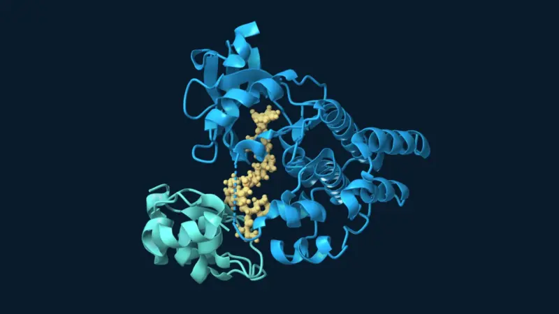
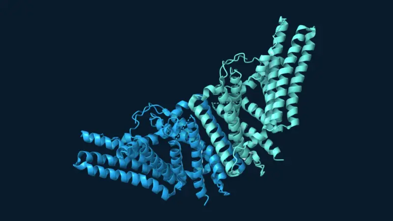
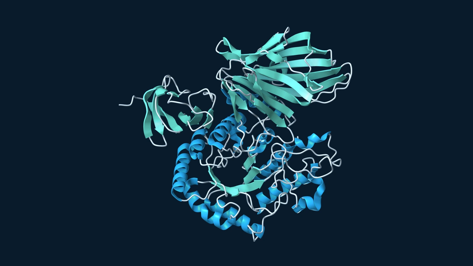
The researchers used two enzymes from the gut bacterium Flavonifractor plautii: FpGalNAc deacetylase and FpGalactosaminidase. Acting in sequence, they modify and remove the terminal sugar that distinguishes the A antigen, exposing the underlying H antigen found in blood group O. The treatment changes these surface carbohydrates; it does not rewrite the kidney’s genes. Original study.
Testing kidneys outside the body
The kidneys came from nine deceased donors. Within each pair, one organ received enzymes and the other served as an untreated control. A machine circulated preservation fluid through the vessels to deliver the treatment.
The team tested both warm perfusion, at approximately 37°C, and cold perfusion, at approximately 4°C. Measurements showed an approximately 80% reduction in A-antigen signal after about two hours with either approach. Cold perfusion was particularly relevant because it was already used to preserve donor kidneys, offering a possible route for treatment during organ preparation or transport.
Across the short experiments, the researchers detected no additional treatment-related injury in the measurements they assessed. However, a small study finding no detectable difference cannot establish that the procedure is harmless under every condition. Original study.
An immune challenge, with important limits
Three treated kidneys and their paired controls then underwent a four-hour warm-perfusion challenge using fluid containing blood group antibodies. Treated organs showed minimal anti-A antibody binding and lacked the tissue deposits of activated complement seen in untreated kidneys. Complement is an immune defence system that can contribute to transplant injury.
This was a laboratory model of an incompatible transplant. The circulating fluid lacked platelets and clotting factors, leaving out an important part of the rejection process. The experiment also could not determine what would happen after the kidney began making A antigens again. Original study.
Calling the result a “universal donor kidney” therefore needs care. Removing A antigens addresses one compatibility barrier. Transplant matching also considers tissue type and antibodies against the donor, as Mayo Clinic’s overview explains. Blood group conversion would not make every kidney suitable for every recipient.
Later evidence: update added 21 September 2026
A separate study published online on 3 October 2025 moved beyond isolated-organ experiments. Jun Zeng and colleagues transplanted an enzyme-converted kidney into a blood group O recipient who had been declared brain-dead. This was research in a human decedent model, not evidence of recovery in a living transplant patient.
The published abstract reports no hyperacute rejection and no evidence of antibody-mediated rejection during the first two days. From day three, however, antibody-mediated lesions and complement deposition appeared as A antigens returned. Those later findings matter: avoiding an immediate immune attack did not demonstrate lasting protection.
The next question is how to preserve the initial compatibility benefit as the organ renews its surface molecules. The evidence described here leaves durable function and safety in living recipients to be established.
Related reading: Nanozyme Helps Collagen Resist Enzymatic Breakdown.
Update note, 21 September 2026: This archived report was revised to identify the experimental models, remove unsupported claims about patient outcomes, and add a separate account of the later decedent-model study. The original publication date has been retained.
References
- MacMillan, S., Hosgood, S. A., Walker-Panse, L., et al. (2024). Enzymatic conversion of human blood group A kidneys to universal blood group O. Nature Communications, 15, 2795. DOI: 10.1038/s41467-024-47131-9.
- Zeng, J., Ma, M., Tao, Z., et al. Enzyme-converted O kidneys allow ABO-incompatible transplantation without hyperacute rejection in a human decedent model. Nature Biomedical Engineering, 10, 1154–1170 (2026); first published online 3 October 2025. DOI: 10.1038/s41551-025-01513-6.
- Mayo Clinic. Kidney transplant: finding a match. Clinical background on blood typing, tissue typing and crossmatching.