Give two versions of a Japanese firefly enzyme the same light-producing substrate. One reaction glows yellow-green; the other glows red. The difference need not be a dye or a pigment sitting in front of the light. A change from serine to asparagine at one position in the enzyme itself can shift the colour.
This makes firefly luciferase a good test of a deceptively simple question. If the reaction starts with the same luciferin, what can the protein change? Researchers have approached the question in two quite different ways. They have measured how a pocket fits around a molecule. And they have counted photons across the spectrum to see what it actually means for light to become redder.
Which molecule emits the light?
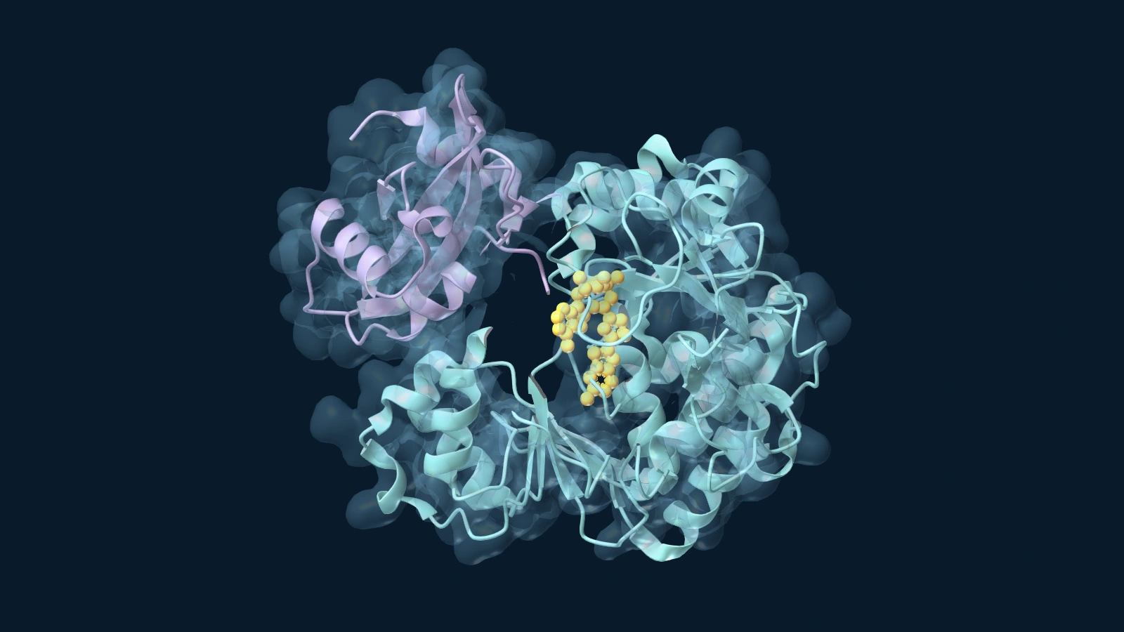
Luciferin is the starting substance; luciferase is the enzyme. Using ATP in the presence of magnesium, the enzyme activates luciferin by attaching an AMP group. The activated molecule reacts with oxygen, and the chemistry produces oxyluciferin in an electronically excited state. When an emitting molecule settles into a lower-energy state, it can release energy as a photon. A red photon has less energy than a green one.
ATP helps set up the chemistry. It is not itself the luminous substance, any more than luciferase is a tiny piece of glowing glass. This reaction provides an instructive contrast with how ATP synthase makes ATP: one enzyme helps supply ATP, the other uses it to activate a substrate. The light-producing reaction also differs from fluorescence, in which an incoming photon first excites the material. Firefly bioluminescence supplies that excitation through chemistry.
The energy of an electronic transition depends on more than the molecule’s skeleton. Charges, nearby groups and the freedom to move can all affect how an excited molecule behaves. Changing an amino acid in the pocket can therefore alter the distribution of emitted wavelengths without changing which luciferin went into the reaction.
The experiment that found a difference in fit
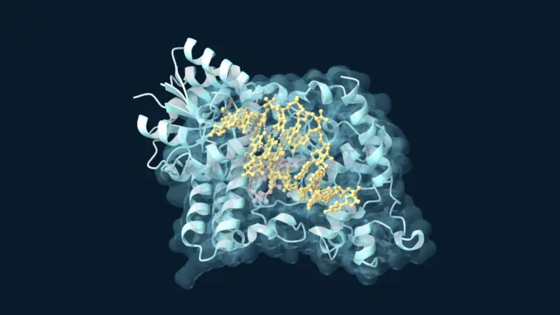
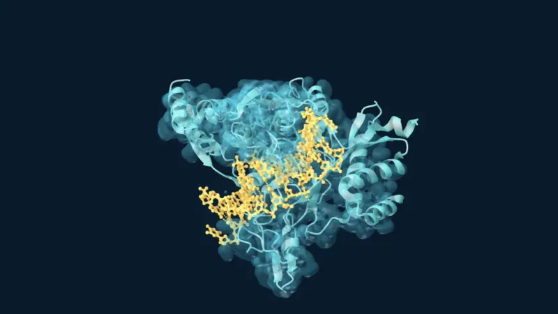
Toru Nakatsu and colleagues investigated a luciferase from the Japanese Genji firefly. Their structural comparison included a red-emitting version in which asparagine replaced serine at position 286. The obstacle was that an excited light emitter does not wait around to be examined by X-ray crystallography. The team used a stable analogue, DLSA, shaped to stand in for an intermediate. That made it possible to compare pockets occupied by the same compound.
Look at the red-emitting variant and its comparison structure. The mutant’s overall fold is recognisable. What interested the researchers was a local change involving a neighbouring amino acid, isoleucine 288. Comparisons across complexes suggested that in the reference enzyme this side chain could move in towards one end of the bound molecule. The same close fit was not seen in the red mutant. The researchers proposed that differences in how tightly the protein constrains the emitter help select a colour.
Neither amino acid is a coloured filter. Nor is the analogue in those coordinates emitting the flash that was measured in a separate reaction. A crystal structure tells us where atoms can fit. A spectrum tells us what light comes out. Putting those measurements together suggested a mechanism; it did not turn the X-ray image into a photograph of excited oxyluciferin.
Redder does not necessarily mean more red
There is another way to change what a viewer sees. Imagine a lamp emitting both green and red light at once. If the green output fades while the red holds steady, the lamp looks redder. No part of its output has had to slide continuously from green to red. A camera photograph alone would not distinguish that change from one in which red output actually increased.
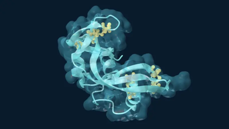
In the measurements across pH, Yoriko Ando and colleagues measured intensities as a function of wavelength. They found that a pH-sensitive green contribution was particularly important for the colour change in the firefly reaction they studied. In the later mutation study, researchers compared the spectra of a Japanese firefly luciferase with variants altered at a different position, 257. Changing that position changed the relative strength of the green emission substantially; the longer-wavelength contributions were comparatively stable in several of the conditions tested.
Those latter results are not another measurement of the 286 mutant. They are an independent reason to be careful about the word “shift”. A broad spectrum can change shape because different contributions change in strength. Fitting a spectrum with several mathematical peaks is useful, but does not by itself prove there are exactly that many distinct chemical species in the active site. A separate time-resolved optical study found that assigning pH-dependent colour to a single species would oversimplify the chemistry.
This is a subtler example of why a protein’s arrangement matters to activity. The enzyme can still complete the reaction after a small structural change, yet the light gives away a difference. For an experimenter using the glow as a signal, that matters: a detector counting only green photons can report a weaker signal even when longer-wavelength photons are still arriving. To find out why a firefly reaction looks redder, the first useful question is not simply “where is the red pigment?” It is “which part of the spectrum changed?”