You can sit perfectly still and still be running an immense energy operation. Cells must maintain their chemical balance, move materials and keep building the molecules they need. Much of that work depends on a molecule called ATP—and on a remarkable enzyme that helps replenish it.
ATP synthase does something that sounds almost too mechanical for biology: part of it rotates. A flow of tiny charged particles drives a molecular rotor, and that motion helps the enzyme make ATP.
Long before CRISPR became a household name, this enzyme was at the centre of a Nobel Prize story about how living things turn energy into usable chemical work.
Your cells keep recycling their spending money
ATP stands for adenosine triphosphate. Its three phosphate groups make it part of a chemical system that cells can couple to many jobs. When ATP is converted to ADP and phosphate, the overall reaction can help power otherwise unfavourable processes.
Calling ATP the cell’s currency is useful: different activities can draw on the same chemical payment system. But the currency must be replenished. ATP synthase adds phosphate to ADP, making ATP again. It transfers energy into a usable form; it does not create energy from nothing.
In our mitochondria, the cell’s energy-converting compartments, this process is linked to the breakdown of food. Related ATP synthases work in bacteria and in the chloroplasts where plants capture sunlight. The surroundings and energy supply differ, while the central problem remains familiar: how can a cell turn an available energy source into ATP?
A membrane makes the machinery possible
A membrane can separate two chemical environments. In mitochondria, other molecular machines use energy from electron transfers to move protons—hydrogen ions—across the inner membrane. The result is a difference in both charge and proton concentration.
That difference is an energy store. Imagine water held behind a dam: allowing it to return through machinery can do useful work. In a cell there is no waterfall. Charged particles and molecular interactions replace the moving water and large metal turbine. The analogy explains the direction of energy flow, rather than every detail of the mechanism.
This membrane-based explanation grew from Peter Mitchell’s chemiosmotic theory, recognized by the 1978 Chemistry Nobel Prize. It supplied essential context for another question: how did the enzyme actually use that stored energy?
The rotating part changes the chemistry
ATP synthase has a membrane-embedded section, called Fo, connected to a projecting catalytic head, F1. Protons moving through the membrane section can drive rotation of a ring and central shaft. A supporting stalk helps keep other parts in place, so that the whole assembly does not simply turn together.
The head contains three catalytic sites. They change shape in a coordinated sequence, binding ingredients, forming tightly held ATP and releasing the product. Paul D. Boyer’s binding-change mechanism made these changes in binding central to the explanation. A major role of the energy input is to alter how tightly the enzyme holds its molecules, including enabling ATP to leave.
Picture three hands taking turns opening and closing around their contents. The shaft coordinates the changes; it does not hammer phosphate onto ADP. Nor are the hands rigid mechanical claws. The real machine is a flexible assembly of proteins whose changing shapes affect chemical reactions.
Watch the molecular motor at work
A crystal structure made the proposal visible
In 1994, John E. Walker and colleagues reported an X-ray structure of the F1 portion from bovine heart mitochondria. The three catalytic beta subunits were different in shape and in the nucleotide molecules they held. Rather than three identical sites frozen at the same stage, they resembled different moments in a repeating cycle.
That structure supported the binding-change explanation. It was not a complete picture of the membrane motor, and a crystal could not itself show the shaft spinning. It provided a physical arrangement that experiments could test.
The 1997 Chemistry Nobel Prize gave half the award jointly to Boyer and Walker for explaining the enzymatic mechanism of ATP synthesis. The other half went to Jens C. Skou for discovering the sodium–potassium pump, a different enzyme that transports ions using ATP.
Then researchers watched a tiny marker turn
Another experiment made the rotation wonderfully tangible. Hiroyuki Noji, Ryohei Yasuda, Masasuke Yoshida and Kazuhiko Kinosita Jr attached a fluorescent actin filament to the central shaft of an immobilized F1 motor. The filament acted as a large visible pointer on a molecular axle.
Their 1997 paper reported watching that pointer rotate when ATP was supplied. The isolated head was consuming ATP to drive motion—the reverse direction of energy conversion from the ATP-making operation described above.
In a 2004 experiment, researchers used an attached magnetic bead to force the shaft around in the appropriate direction and detected newly made ATP. The enzyme’s reversibility had become something they could manipulate, linking mechanical motion directly to chemical production.
The machine is flexible, and the pictures have limits
Today, researchers can compare structural snapshots of more of the assembly. A 2020 cryo-electron microscopy study of the bacterial enzyme revealed several rotational substates and flexing that helps its coupled motors work together.
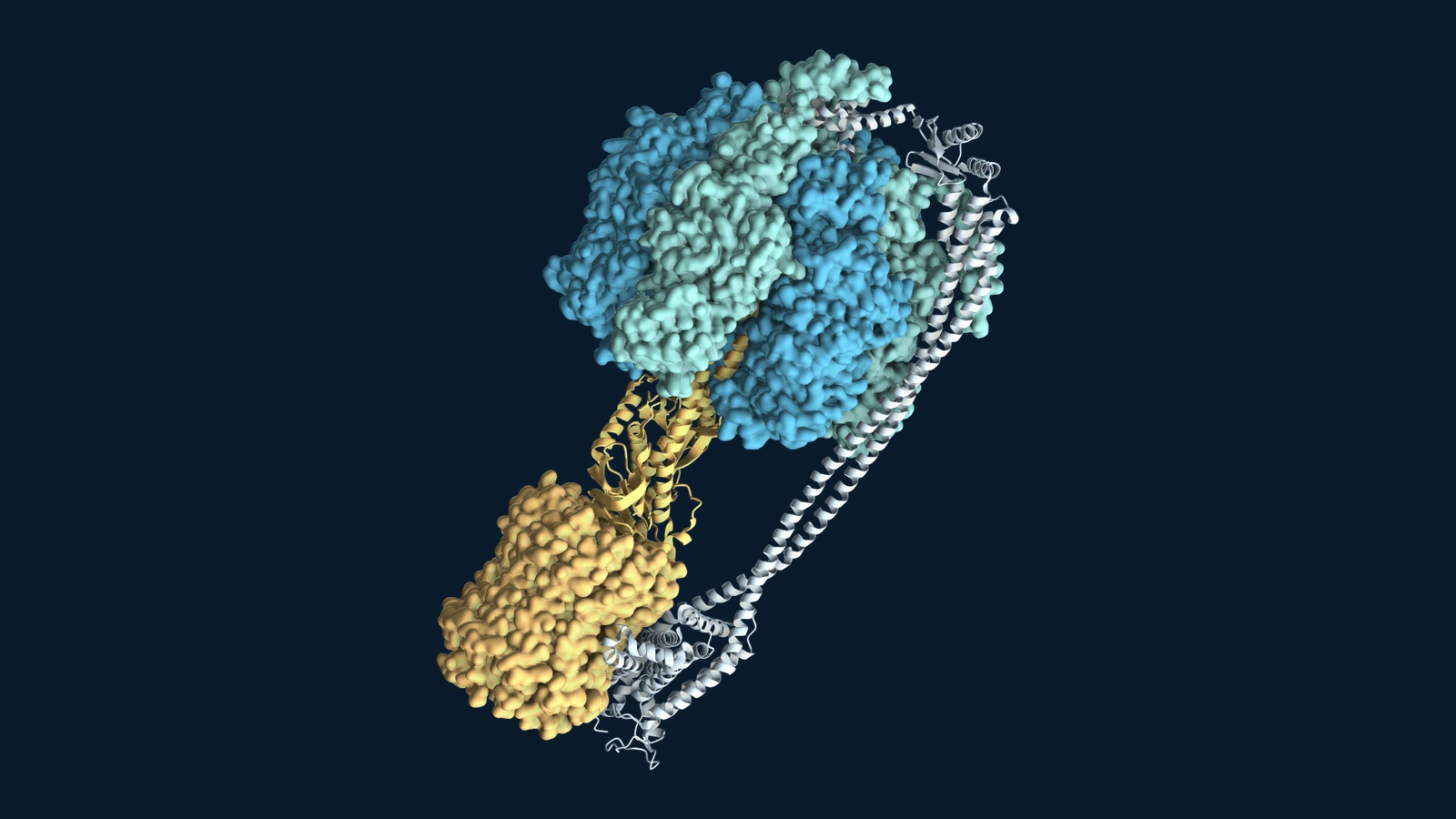
The Mol* portrait accompanying this article uses one experimental structure of the Escherichia coli complex. It shows the membrane section as well as the catalytic head. It is not the human enzyme, and the coordinates capture an inhibited laboratory state, rather than a running motor. Colour helps distinguish components; it does not show their natural appearance.
ATP synthase is also not the only route to ATP. Cells can make ATP through other reactions, and the balance depends on the organism and circumstances. Its importance rests on the enormous range of life that uses this particular conversion between a membrane gradient, rotation and chemistry.
Explore the wider history of enzymes and the Nobel Prizes, and ATP synthase stands out for a simple reason: the molecular world contains working rotary machinery. The remarkable part is how a turning protein helps keep living chemistry supplied.
Sources and further reading
- NCBI Bookshelf: The mitochondrion, from Molecular Biology of the Cell, on gradients and ATP synthase.
- NobelPrize.org: The 1978 Chemistry Nobel Prize, awarded to Peter Mitchell.
- NobelPrize.org: The 1997 Chemistry Nobel Prize, including the exact division of the award.
- Abrahams and colleagues (1994): Structure at 2.8 Å resolution of F1-ATPase from bovine heart mitochondria.
- Noji and colleagues (1997): Direct observation of the rotation of F1-ATPase.
- Itoh and colleagues (2004): Mechanically driven ATP synthesis by F1-ATPase.
- Sobti and colleagues (2020): Cryo-EM structures of E. coli ATP synthase.