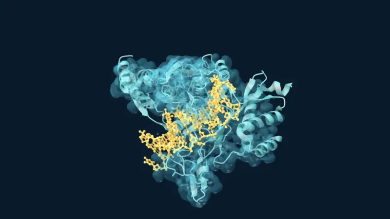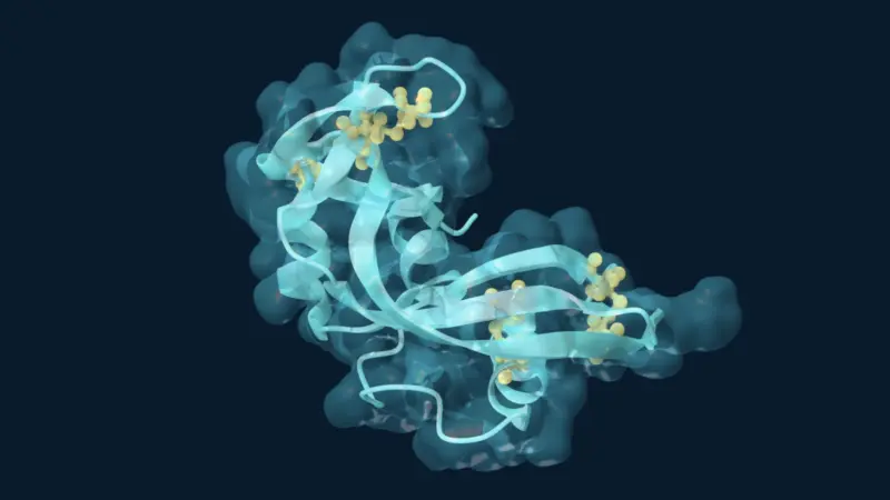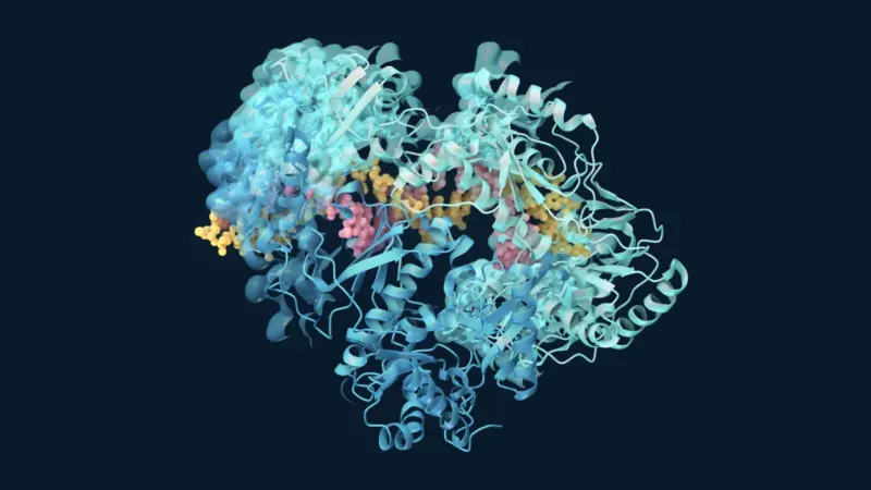
An enzyme involved in metabolism can also build striking patterns of triangles within triangles. In research published in Nature on 10 April 2024, Franziska L. Sendker and colleagues described this behaviour in citrate synthase from the cyanobacterium Synechococcus elongatus. The discovery adds an unusual geometry to the known ways natural proteins assemble.
A triangle built from smaller triangles
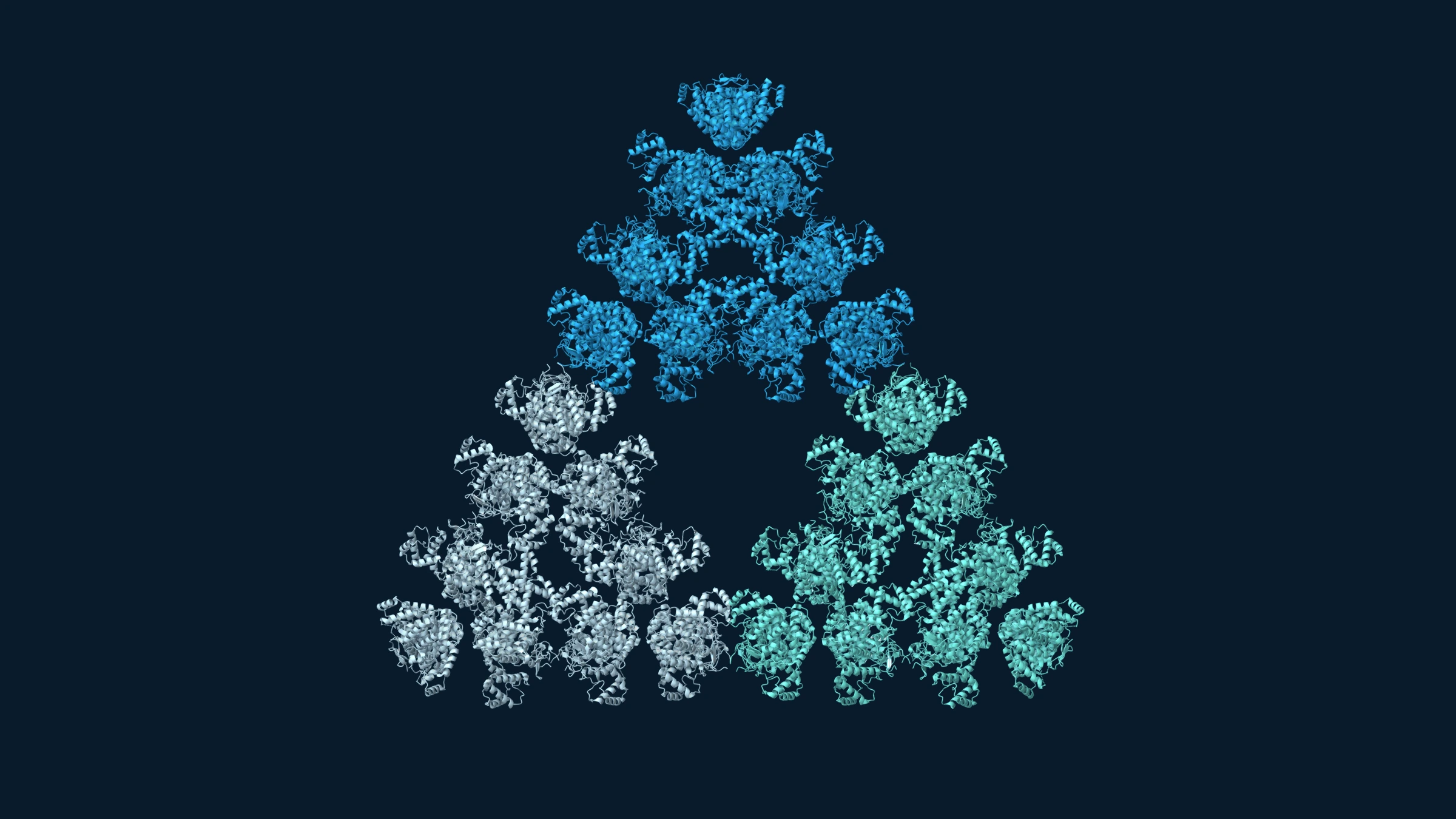
The building block contains six enzyme subunits: a hexamer. Three hexamers form an 18-subunit triangle, and three of those form a larger, 54-subunit triangle. The smaller triangular arrangements recur within the larger one, leaving openings between them. The deposited structures include the wild-type 18-subunit assembly and a 54-subunit model of the H369R variant.
These are finite molecular versions of a Sierpiński triangle. The researchers also detected larger assemblies, but did not establish that structures beyond 54 subunits retained the same fractal geometry. An infinitely repeating mathematical fractal is an idealization, not an experimentally observed molecule. Original study.
How the pattern emerges
Cryo-electron microscopy helped reveal why the protein forms these open triangles. Neighbouring chains do not all make equivalent contacts. Small differences in their arrangements allow connections at particular positions while preventing other positions from filling in. This preserves the gaps that distinguish the fractal pattern from an ordinary molecular lattice. Max Planck research account.
Effects on activity, but an uncertain biological role
In laboratory assays, fractal assembly reduced citrate synthase activity. Adding substrates or the product promoted disassembly into hexamers; pH also influenced assembly. Thus, the structures had measurable biochemical effects.
However, bacteria carrying a variant that could not form fractals showed no detected growth difference under continuous light or 12-hour light–dark cycles. Recovery after nitrogen starvation was also similar. These results do not exclude a role under other conditions. They distinguish effects on purified enzyme activity from a demonstrated benefit to the organism. Study results.
Could the fractal be an evolutionary byproduct?
The team inferred ancestral protein sequences and produced the reconstructed enzymes to investigate how the assembly arose. This approach provides experimentally testable models of evolutionary history; it does not recover ancient proteins directly. The authors’ institutional account describes an assembly that appeared through few sequence changes and was subsequently lost in related lineages.
In one reconstructed ancestral background, a single amino-acid substitution enabled fractal assembly. The same change did not work in more distant ancestors, showing that the surrounding sequence mattered. Together with the physiological tests, this supports—but does not prove—an evolutionary origin without selection for the fractal itself. Original study.
Why the finding matters
For enzyme research, the useful distinction is between three questions: what a protein assembly looks like, how it changes catalysis, and whether that change helps the organism. This study connects all three, while leaving the last question open. Its intricate geometry makes a compelling image; the measured effects and remaining uncertainties make the scientific story.
Related reading: AlphaFold 3: Predicting How Biomolecules Fit Together.
Update note, 20 September 2026: This archived report was revised to distinguish finite molecular assemblies from an ideal mathematical fractal, describe the tested biological conditions, and clarify the limits of the evolutionary interpretation. The accompanying image was replaced with a new rendering of deposited structural data.
References
- Sendker, F. L., Lo, Y. K., Heimerl, T., et al. (2024). Emergence of fractal geometries in the evolution of a metabolic enzyme. Nature, 628, 894–900. DOI: 10.1038/s41586-024-07287-2. Open-access full text.
- Max Planck Society (10 April 2024). Discovery of the first fractal molecule in nature.
Image sources
- Structural coordinates: RCSB PDB 8RJK, the cryo-EM-based model of a 54-subunit assembly of the H369R citrate synthase variant, described by Sendker et al. (2024).
- Visualization: Sehnal, D., et al. (2021). Mol* Viewer: modern web app for 3D visualization and analysis of large biomolecular structures. Nucleic Acids Research, 49, W431–W437. DOI: 10.1093/nar/gkab314.