A strand of three amino acids enters the fungal enzyme isopenicillin N synthase. What emerges is hardly recognisable as a strand: two rings now share an edge. One is the four-membered β-lactam ring that gives penicillins their family resemblance. The chemistry looked plausible on paper for decades. The hard part was finding out which atoms were touching between the beginning and the end.
In October 2026, Patrick Rabe and colleagues reported an unexpected separation in the middle of that sequence. The substrate initially holds on to the enzyme’s iron atom through a sulfur atom. For a short interval, that connection breaks. When the sulfur comes back to the iron, there is already a ring. The time-resolved study makes a useful point about watching enzymes work: sometimes the most informative event is the disappearance of a bond, rather than the formation of one.
How to catch half a penicillin
The enzyme, isopenicillin N synthase, works on a tripeptide known as ACV. It uses iron and molecular oxygen to turn ACV into isopenicillin N. The first ring is the β-lactam; the second is a five-membered ring containing sulfur. The product is a starting point for making other antibiotics. This research concerns how that core is built. It does not test an antibiotic against an infection.
A structure taken before the reaction will tell you where to look. A structure taken afterwards will tell you where the atoms ended up. Neither establishes which path they followed. The earlier experiments with a shortened enzyme provided chemical evidence for a fleeting sulfur-containing species called a thioaldehyde. They did not settle how that species sits relative to the metal in the intact working enzyme.
The team gave themselves two ways to catch it. First, they prepared microscopic crystals containing enzyme, iron and peptide without oxygen. Introducing oxygen provided a starting signal. An X-ray free-electron laser could then collect diffraction from different crystals after different delays. That distinction matters: each snapshot combines measurements across crystals. No camera follows an individual substrate from entry to exit.
The second trick was to replace a particular hydrogen atom on the peptide with deuterium, a heavier isotope. Breaking the relevant bond becomes slower. This holds up a later step, giving the preceding chemical species time to accumulate. The labelled substrate still produces the ring system, but the experimental clock is deliberately different. The seconds attached to the structures are exposure times in this arrangement, not a timetable for every enzyme in a fungus.
What changed around the iron in isopenicillin N synthase
In the starting structure, the peptide’s cysteine sulfur is coordinated to iron. Early oxygen-exposed samples place oxygen at the metal. A few seconds further into the series, the data support a thioaldehyde in which sulfur no longer coordinates the iron. That is a surprisingly specific answer: a depiction that leaves the sulfur attached throughout would miss precisely what the experiment resolved.
Later samples show sulfur coordinated again, this time in a species with the β-lactam ring already formed. There is a subtlety visible in the eight-second structure. The diffraction data fit a mixture of states. A time label on a coordinate file does not mean every enzyme molecule in that sample had reached the same step. At one ten-second time point the researchers found evidence for some completed product alongside the one-ring intermediate. The later product structure gives a clearer view of the destination.
Assigning an iron oxidation state poses a different problem from placing sulfur. The shape of an electron-density map alone is not a label for the iron’s electronic state. The researchers paired diffraction with X-ray emission spectroscopy and tested the effects of replacing specific amino acids. Together with calculations, these results support a pathway involving a highly oxidised iron centre and water molecules in the active site. The observations constrain that interpretation; they are not photographs of protons passing from atom to atom.
What a molecular movie can honestly show
In EnzymeNews’s ATP synthase film, motion connects experimentally determined structures. Here the researchers went a step further experimentally: by starting the reaction and waiting known intervals, they could establish an order among changes in the crystal populations. The clever substitution of deuterium improved the view of chemistry that would otherwise proceed too quickly. It also matters when interpreting the pace of the result.
Watch the sulfur atom, shown in gold. The film opens as it bonds to carbon and closes the second ring of isopenicillin N, then rewinds to the open chain. As the reaction unfolds, look for the moment sulfur loses contact with iron. X-ray snapshots show where sulfur sits at key stages. Simulations of the enzyme’s active site show how the two ring-closing bonds could form.
Molecular models and credits
The enzyme is isopenicillin N synthase from the fungus Aspergillus nidulans. It uses an iron ion and molecular oxygen to turn the peptide ACV (δ-(L-α-aminoadipoyl)-L-cysteinyl-D-valine) into isopenicillin N. In the crystal experiment, replacing one hydrogen on valine with deuterium gave the labelled substrate ACdV. The film depicts ordinary hydrogen during the simulated transfers.
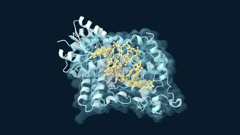
- 8PY6 supplies the article image of the starting enzyme with iron and the labelled peptide, before oxygen exposure.
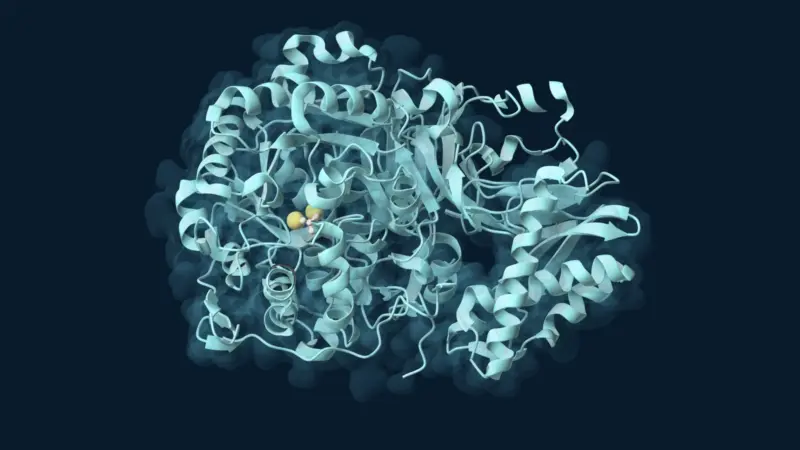
- 8PY7 supplies the enzyme structure used throughout the film and shows the iron, oxygen and peptide in its active site.
- 8PYA contains two modelled states, the starting peptide and a thioaldehyde intermediate (IO0). Their sulfur atoms occupy different positions relative to iron.
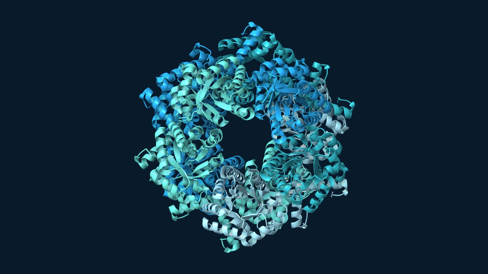
- 9I3B supplies the crystal packing and the electron-density map around the trapped, one-ring β-lactam (A1IZG). The crystal contains a mixture of states, including the modelled iron–oxo species.
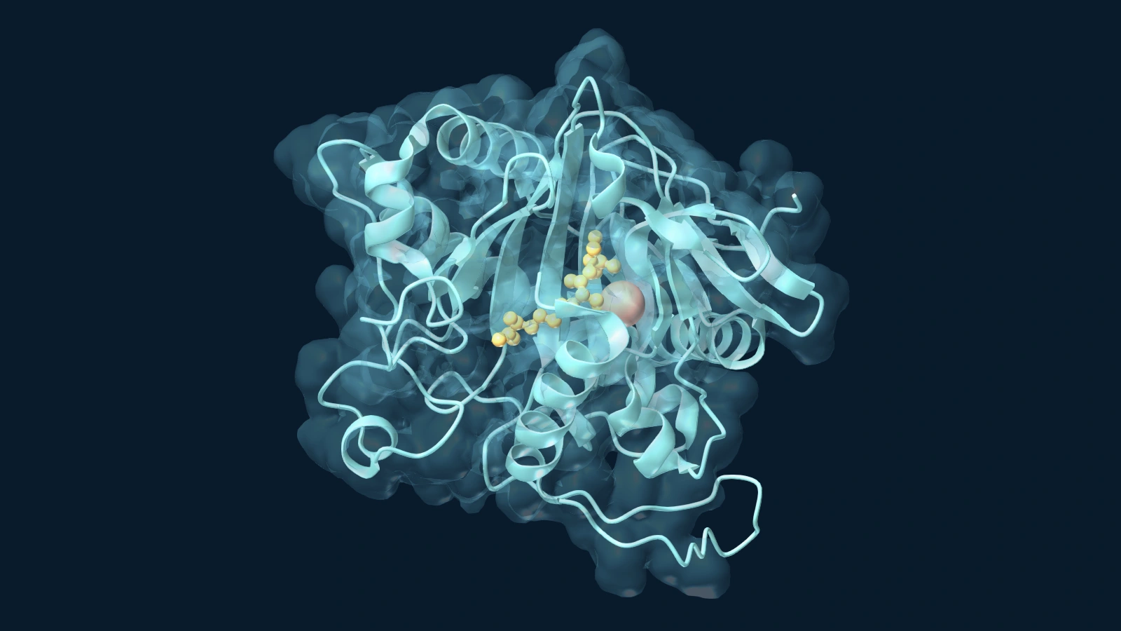
- 9QPH contains isopenicillin N (IP1). Its two joined rings provided the experimental reference for the finished product.
Additional structures examined during production were 8PY9, 9I3A, 9I3C and 9I3D. Patrick Rabe and colleagues deposited the structures and published the study and supporting coordinates. EnzymeNews aligned and adapted selected coordinates under CC BY 4.0 for the local simulations of the two ring closures.
There is a useful lesson in why this experiment took more than better cameras. The crucial design decision was chemical: delay the second ring long enough to see evidence of the first. Without that choice, a perfectly sharp picture of the final antibiotic could leave the most interesting question unanswered. The lasting result is a narrower, much more testable account of the pathway: sulfur need not stay attached to the metal while the enzyme turns a straight chain into a ring.