It is easy to imagine a Nobel Prize beginning with a telescope, a particle accelerator or an extraordinary new medicine. This one began with a bean.
Inside the jack bean was urease, an enzyme that breaks down urea. In 1926, James B. Sumner turned it into tiny crystals and challenged a basic uncertainty about life: what, exactly, were enzymes made of? A substance associated with living things could be isolated, tested and handled as a chemical material.
A century later, urease still connects apparently distant worlds: the protein bottle on a laboratory shelf, the nitrogen in a farmer’s field and the long effort to understand how biological molecules work.
A small reaction with a large footprint
Urea is a small molecule containing nitrogen. Urease uses water to help break it down, producing ammonia and carbon dioxide overall. In water, the ammonia can pick up hydrogen ions to become ammonium; the balance depends on the surrounding conditions. That chemistry can also make the local environment more alkaline.
The enzyme supplies a faster route through the reaction. It does not manufacture nitrogen or use itself up as an ingredient. Think of it as a carefully shaped workbench: the right materials meet in a setting that makes a difficult rearrangement easier. The analogy has limits. There is no worker making decisions; the arrangement and chemical properties of atoms do the work.
Plants, some bacteria and fungi make ureases. The EMBL-EBI Mechanism and Catalytic Site Atlas describes their role in helping organisms use urea as a nitrogen source. Their shared reaction does not mean that every organism packages or uses the enzyme in exactly the same way.
The bean that became an argument
Sumner chose jack beans for practical reasons. They contained plenty of urease, and its activity could be measured by tracking the ammonia formed from urea. He had a chemical signal to follow while separating the enzyme from everything else in the bean.
His 1926 paper describes small, colourless crystals whose dissolved material had strong urease activity and gave chemical tests characteristic of protein. The result followed almost nine years of work, rather than one lucky afternoon.
Urease became the first enzyme successfully crystallized. The crucial claim was that the protein was the enzyme itself. Seeing crystals was not enough: scientists also needed evidence linking the material in those crystals to the reaction. Otherwise, an active trace contaminant might have been doing the chemistry while the visible protein merely carried it along.
Why beautiful crystals did not settle everything
That alternative became a serious objection. As the Nobel Foundation’s historical account explains, some researchers doubted that Sumner had isolated the active substance. Could the enzyme have been trapped in, or attached to, another crystalline material?
Purification is a demanding kind of detective work. If two substances repeatedly travel together, that is evidence of a connection, but it does not automatically establish which one causes an effect. The debate was about the strength of that connection, not whether the crystals were attractive.
Further work, including John H. Northrop’s crystallization and study of digestive enzymes, helped establish protein enzymes as substances that could be purified and investigated. In 1946, Sumner received half the Chemistry Nobel Prize for showing that enzymes could be crystallized. Northrop and Wendell M. Stanley shared the other half for separate work preparing enzymes and virus proteins in pure form.
Two nickel ions at the working centre
Modern structural biology lets us look far beyond the outline of a crystal. Jack bean urease contains a working pocket with two nickel ions. The surrounding protein holds this metal-containing centre in an arrangement suited to the chemistry.
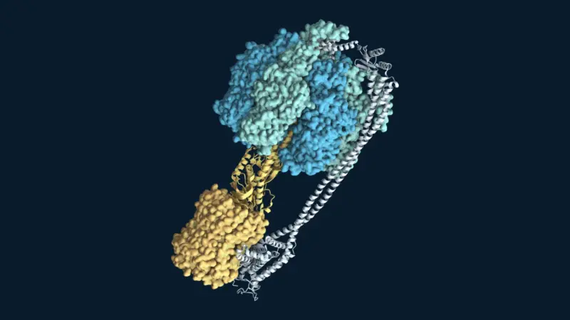
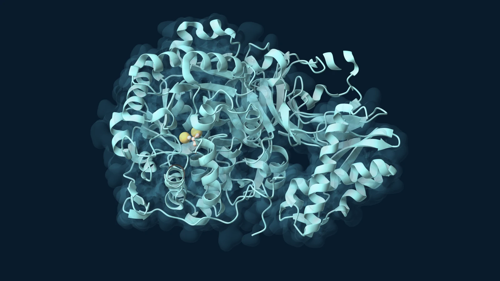
To show how the substrate meets that centre, our molecular portrait uses a later structure of urease from the bacterium Sporosarcina pasteurii. It is a different organism from Sumner’s jack bean, with a different arrangement of protein chains, but it reveals the same type of nickel-containing working pocket. The experimental structure, PDB 6QDY, was reported by Mazzei and colleagues in 2019.
The illustration shows one three-chain catalytic unit from the larger nine-chain enzyme. Aqua ribbons trace the protein’s fold, a transparent surface suggests its outer shape, and gold spheres mark the two nickel ions. The small peach-coloured molecule beside them is urea, placed exactly where the experiment found it. The colours are visual choices, rather than the molecules’ natural appearance.
Capturing urea in this pocket was difficult because the enzyme reacts so quickly. The researchers used fluoride to replace a reactive hydroxide and stop the chemistry after urea had bound. Their structure therefore captures a trapped substrate complex. The illustration shows that fixed arrangement, rather than urea being broken down.
Sometimes the useful move is to slow it down
In agriculture, the speed of urease can become a problem. Urea is widely used as a nitrogen fertilizer. Once it reaches a field, the timing of its conversion matters: nitrogen that leaves as ammonia is unavailable for the crop that the farmer intended to feed.
One response is to use a urease inhibitor, a substance that slows the enzyme’s action. In a USDA Agricultural Research Service field study in Idaho, urea treated with the inhibitor NBPT lost less ammonia than untreated urea left on the soil surface. Incorporating fertilizer into the soil also reduced losses in that experiment.
This does not make an inhibitor a universal answer. The study concerned a particular soil and field setting, and its authors called for testing across more locations and seasons. Product choice, placement and conditions belong to the same story as the enzyme. A fast biological reaction can be valuable in one setting and poorly timed in another.
A familiar laboratory bottle, with an unfamiliar past
Urease makes an invisible chemical event measurable. Follow its products, and you can follow its activity. That was part of Sumner’s advantage, and it remains an inviting way to understand enzyme experiments: ask what changes, how quickly it changes and what happens when the protein is altered.
There is also a useful limit to the historical lesson. Urease helped establish that an enzyme could be a protein; it did not prove that every biological catalyst must be one. The 1989 Chemistry Nobel Prize later recognized discoveries showing that RNA can catalyse reactions too.
For the broader story, see our history of enzymes and the Nobel Prizes. Urease’s own achievement remains striking: a bean protein helped turn a question about life into something scientists could hold, measure and challenge.
Sources and further reading
- Sumner (1926): The isolation and crystallization of the enzyme urease, original paper.
- NobelPrize.org: The development of modern chemistry, including the 1946 award and the enzyme-purity debate.
- Sumner’s Nobel Lecture (1946): The chemical nature of enzymes.
- RCSB PDB 3LA4 and Balasubramanian and Ponnuraj (2010), experimental jack bean urease structure.
- EMBL-EBI Mechanism and Catalytic Site Atlas: Urease, curated reaction and active-site information.
- USDA ARS: Ammonia volatilization from fertilizer sources on a loam soil in Idaho, field research on fertilizer placement and urease inhibition.
- NobelPrize.org: 1989 Chemistry award ceremony speech, on catalytic RNA.
- RCSB PDB 6QDY and Mazzei and colleagues (2019), the bacterial urease–urea complex depicted in this article.