In a laboratory freezer, a tiny tube labelled EcoRI looks thoroughly ordinary. Its contents help researchers cut DNA. The name is so familiar that it can disappear into the background of an experiment, somewhere between the ice bucket and the next coffee.
Yet the ability to make a predictable cut in DNA helped turn genetic engineering from an ambition into something scientists could actually do. Long before CRISPR became a household name, enzymes such as EcoRI gave researchers a way to take genetic material apart and put selected pieces together.
EcoRI also carries a revealing Nobel connection. The 1978 medicine prize recognised the discovery of restriction enzymes and their use in molecular genetics. EcoRI is one of the most recognisable members of that family, although it was not the enzyme at the centre of Hamilton Smith’s original discovery.
A bacterial defence with a second career
EcoRI comes from Escherichia coli, a bacterium better known outside laboratories for its association with our intestines and, in some strains, food poisoning. For the bacterium, the enzyme belongs to a defence system. Viruses that infect bacteria introduce genetic material into their hosts. Cutting incoming DNA can interrupt that invasion.
A dangerous question follows: how does a DNA-cutting enzyme avoid destroying the cell that makes it? In restriction–modification systems, a partner enzyme adds small chemical marks to the bacterium’s own DNA. Those marks help protect it from the matching cutter. The protection depends on chemistry, rather than an enzyme somehow knowing who owns a molecule.
This relationship between destruction and protection helped Werner Arber develop the framework behind restriction enzymes. His Nobel lecture describes how research on bacteria and their viruses opened a route into molecular genetics. A survival mechanism became a research tool because scientists learned to separate the useful cutting activity from its original cellular job.
Six letters that change what happens next
DNA contains four chemical letters, abbreviated A, T, G and C. EcoRI recognises a particular six-letter sequence: GAATTC. It usually cuts between the G and the first A on each strand of the double helix. The two cuts are offset, leaving short, exposed ends rather than a flush break.
Those exposed ends are often called “sticky ends”. The word does not mean they are coated in glue. Their letters can pair with a matching end on another DNA fragment. A separate enzyme, DNA ligase, can then seal the backbone to make a stable joined molecule.
That is an extraordinary change in scale. Researchers cannot handle individual DNA strands with miniature scissors and fingers. EcoRI lets molecular recognition do the positioning. Give it suitable DNA, and the sequence itself determines the places available for cutting.
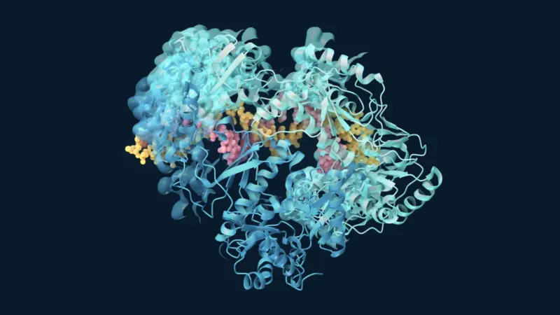
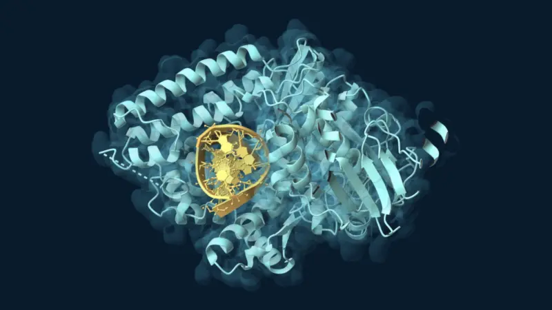
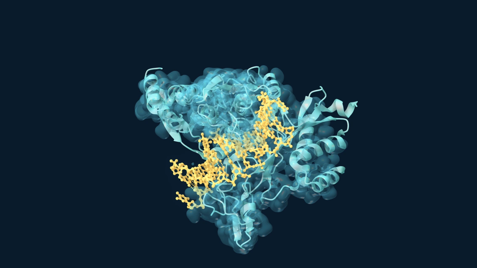
The enzyme works as a pair of matching protein subunits. The experimental EcoRI–DNA structure shows how the protein embraces its target. The helix is a physical object with grooves, surfaces and chemical groups that the enzyme can contact, not merely a string printed on a page.
The remarkable usefulness of a broken end
In 1972, Janet Mertz and Ronald Davis reported a crucial property of the enzyme then called R1 restriction endonuclease: the ends it produced were complementary and could be joined. Their experiments showed why a cut could become the starting point for recombination.
The practical implication was enormous. Fragments from different DNA molecules could carry compatible ends. Those fragments did not need to share an entire history or come from the same original molecule. The chemistry at their edges could make them fit together.
The following year, Stanley Cohen, Annie Chang, Herbert Boyer and Robert Helling constructed functional plasmids by joining restriction-enzyme-generated DNA fragments. Plasmids are relatively small DNA molecules that bacteria can maintain alongside their main chromosome. The reconstructed molecules could function inside bacterial cells and carry properties from their parent DNA molecules.
The achievement went beyond producing an interesting mixture in a tube. A deliberately assembled DNA molecule could survive in a cell and be copied. That helped establish the foundations of DNA cloning: making many copies of a selected piece of genetic material by placing it in a suitable biological carrier.
Where the Nobel Prize belongs
The 1978 Nobel Prize in Physiology or Medicine went to Werner Arber, Daniel Nathans and Hamilton O. Smith. Their contributions connected the biological explanation of restriction, the isolation and characterisation of useful enzymes, and their application to genetic analysis.
Smith and his collaborators worked on an enzyme from Haemophilus influenzae, now known as HindII. Nathans helped demonstrate how restriction enzymes could break a viral genome into reproducible fragments for study. EcoRI belongs alongside that story as a particularly influential tool, rather than as a discovery that should be attributed to the three laureates collectively.
The distinction gives the history more texture. A Nobel Prize marks selected contributions to a larger scientific transformation. Familiar tools on today’s laboratory benches often carry the work of people whose names are absent from the medal’s citation.
Useful does not mean unlimited
EcoRI remains available as a laboratory reagent, including engineered versions. New England Biolabs describes EcoRI-HF as preserving the original recognition sequence while reducing unwanted cutting at alternative sites, a behaviour known as star activity.
The existence of those improvements is a reminder that molecular tools have conditions and compromises. DNA chemistry can affect access to a site, and the wrong environment can reduce specificity. EcoRI also recognises the same short sequence wherever it occurs. It cannot choose one desired occurrence simply because a researcher considers that location important.
For a researcher checking a constructed plasmid, however, a predictable pattern of fragments can be highly informative. A cut becomes a way to ask whether the DNA has the expected arrangement. The same basic property supports both construction and inspection.
A revolution inside an ordinary tube
EcoRI’s lasting appeal is the simplicity of the idea: a protein recognises a small piece of DNA and cuts at a predictable place. From that behaviour came new ways to assemble genetic material and investigate how it works.
Its wider place among enzymes connected with Nobel history is easy to overlook precisely because it became so useful. The little tube stopped looking revolutionary. The revolution became part of the working day.
Sources and further reading
- Nobel Prize in Physiology or Medicine 1978: official award record.
- Werner Arber’s Nobel lecture on restriction and modification.
- Mertz and Davis (1972): EcoRI cleavage generates cohesive ends.
- Cohen and colleagues (1973): construction of functional bacterial plasmids.
- Hamilton Smith’s Nobel lecture on sequence specificity.
- RCSB PDB 1ERI: experimental EcoRI–DNA recognition complex.
- New England Biolabs: engineered EcoRI-HF and specificity.