Pick a carbon atom that has just left a working muscle as carbon dioxide. If you could check its identity again halfway to the lungs, there is a good chance it would no longer belong to a molecule of CO₂. Much of that carbon travels in the blood as bicarbonate. By the time it leaves in an out-breath it is CO₂ again. Why change its form twice?
The distinction matters because a gas and a dissolved ion do different jobs. Dissolved CO₂ can move down a difference in partial pressure, from tissue into blood or from blood into the air in the lungs. Bicarbonate carries carbon in a form that can accumulate in the plasma while the circulation takes it elsewhere. An enzyme inside red blood cells helps move carbon quickly between these two forms. It does not carry the carbon from muscle to lung itself.
Three jobs in a red blood cell
CO₂ enters a red cell from the tissues and reacts with water. The overall products are bicarbonate and a proton. This reaction can occur spontaneously, but carbonic anhydrases speed it enough to contribute during a red cell’s short passage through a capillary. Haemoglobin buffers many of the protons. Meanwhile a membrane protein exchanges bicarbonate for chloride; bicarbonate can leave the red cell and be carried in the surrounding plasma.
Follow that sequence and a common misconception falls away. The carbonic anhydrase is a soluble catalyst; the separate exchanger moves ions across the membrane. Neither of those jobs is accomplished by attaching bicarbonate to haemoglobin. Some carbon does travel bound to proteins, including haemoglobin, and some stays dissolved as CO₂. Bicarbonate is the major route, not the only one. A review of the physiology of carbon dioxide transport sets out the differences.
It is possible to separate the reactions in the laboratory too. In human red-cell exchange experiments, investigators rapidly mixed red-cell suspensions and tracked changes in extracellular pH. Under conditions chosen to make bicarbonate–chloride exchange limit the overall rate, the measured signal reported on transport across the membrane. A pH reading from a suspension is not automatically a stopwatch for a single enzyme; what it tells you depends on which part of the sequence is slow under those conditions.
A gas caught just before it reacts
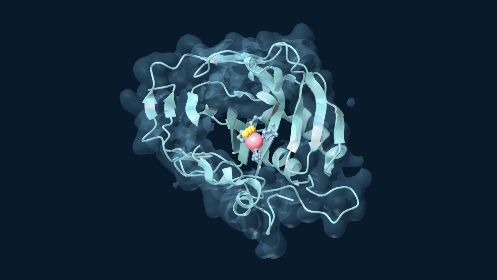
Human red cells contain more than one carbonic anhydrase. Isoform II, a particularly fast one, supplies a beautifully clear view of what catalysis involves. At the bottom of its pocket sits a zinc ion. Zinc helps make a bound water molecule ready to lose a proton. The resulting hydroxide can attack the carbon in CO₂ to form bicarbonate. Product leaves, water returns and the site must be reset for the next molecule.
Speed creates a difficulty for anyone hoping to see the input: the very protein you want to photograph is exceptionally good at making it disappear. In the pressure-trapping experiment, John Domsic and colleagues exposed enzyme crystals to CO₂ under pressure and cooled them rapidly. The zinc-containing structure places the substrate in a largely water-repelling pocket beside the metal.
The researchers also examined the zinc-free comparison. CO₂ could still occupy much the same position without zinc. Capturing and orienting a substrate is one task; helping it react is another. The protein pocket does much of the positioning. The presence of the gas in both structures does not mean that the zinc-free protein can catalyse hydration at the normal rate. Nor does this crystal experiment reproduce the pressures or temperatures inside a red cell.
A proton needs a way out
After bicarbonate departs and water takes its place, the enzyme has another job: transfer a proton from the active-site water towards the surrounding solution. A histidine at position 64 helps with that transfer. To test its role, researchers replaced it with alanine, which cannot shuttle protons in the same manner. In the proton-transfer experiments, the effect depended on precisely what was measured and on the buffer present. Certain imidazole-based buffers could compensate for part of the missing function.
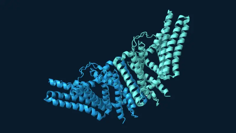
That is a revealing result. If a substance in solution can partly substitute for the removed side chain, the slow step tells you something about how a proton normally gets out. The active-site water study also points to rearrangements of water during product release. Unlike the large rotating parts of ATP synthase, the small changes that matter here can be easy to overlook in an image showing the whole enzyme.
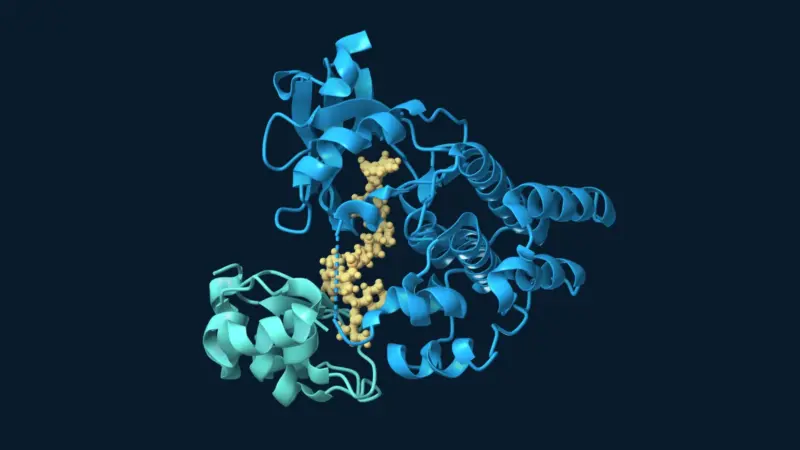
Metal-containing enzymes are not interchangeable. In urease’s nickel-containing active site, the substrate and reaction differ. The identity of the metal, the surrounding protein and the network of water molecules together determine what takes place. Simply finding zinc in a structure would not tell you that the protein helps carry CO₂ through the bloodstream.
At the lung, the same catalyst faces the other way
Air in the lung provides an outlet for CO₂. As CO₂ leaves the blood, bicarbonate enters red cells from the plasma in exchange for chloride. Bicarbonate and a proton can be converted back to water and CO₂, and the gas can diffuse into the air to be exhaled. No second switch has to turn carbonic anhydrase into a different enzyme. The local conditions determine which direction has the greater net flow; the enzyme accelerates movement towards the same chemical equilibrium.
That is why both ends of the journey matter. An enzyme that only sped conversion into bicarbonate might sound useful near muscle, but it would leave the lungs with the wrong cargo to exhale. The same catalyst helps solve both problems. What distinguishes muscle from lung is where the CO₂ is coming from and where it can go next.
Sources and further reading
- Arthurs and Sudhakar (2005). Carbon dioxide transport.
- Chow et al. (1976). Kinetics of bicarbonate-chloride exchange across the human red blood cell membrane.
- Domsic et al. (2008). Entrapment of Carbon Dioxide in the Active Site of Carbonic Anhydrase II.
- PDB 3D92 — zinc-containing structure.
- PDB 3D93 — zinc-free comparison.
- Tu et al. (1989). Role of histidine 64 in the catalytic mechanism of human carbonic anhydrase II studied with a site-specific mutant.
- Kim et al. (2025). Fast product release requires active-site water dynamics in carbonic anhydrase.
- urease’s nickel-containing active site
- ATP synthase
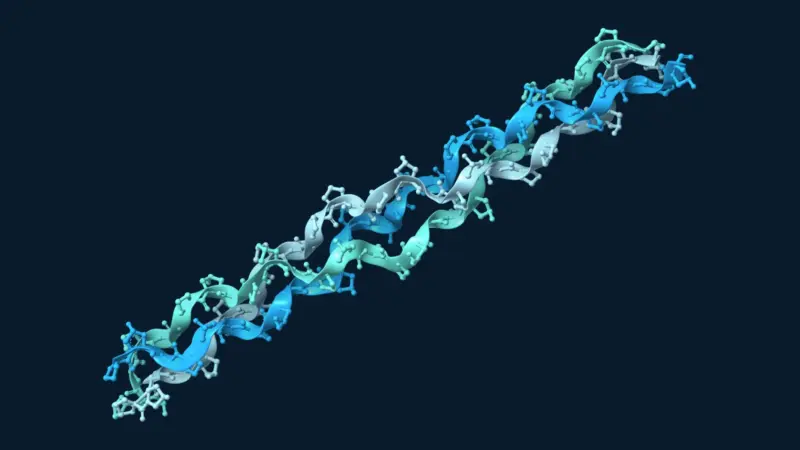
Molecular visualization. EnzymeNews created the article artwork with Mol*. Software reference: Sehnal et al. (2021), Mol* Viewer: modern web app for 3D visualization and analysis of large biomolecular structures, Nucleic Acids Research 49, W431–W437.