A small bottle labelled RNase A can look unremarkable on a laboratory bench. Its contents cut up RNA, a useful job when researchers want to clean RNA out of a DNA preparation. Yet this familiar reagent helped answer one of biology’s deepest questions: how does a long chain of chemical building blocks become a working molecular machine?
The answer mattered far beyond the enzyme itself. Proteins need particular three-dimensional arrangements to do their jobs. A list of ingredients is not enough to explain a finished machine. Where does the instruction for its shape come from?
Ribonuclease A, usually shortened to RNase A, helped turn that question into an experiment. The work led to the 1972 Nobel Prize in Chemistry, shared by Christian Anfinsen, Stanford Moore and William Stein for complementary discoveries about this one compact protein.
A tiny pair of molecular scissors
RNase A’s natural setting is digestion. The pancreatic enzyme helps break down RNA, including RNA in food. The version studied so intensively by biochemists came from cattle. Its chain contains just 124 amino acids, the linked building blocks from which proteins are made.
RNA is a chain too. It carries a sequence written with four chemical letters, usually abbreviated A, U, G and C. RNase A cuts within accessible RNA strands, favouring positions after C and U. Its folded surface includes a groove where part of the RNA can sit near the chemical groups that promote cutting.
Calling it scissors is useful, provided we do not imagine miniature metal blades. The enzyme helps rearrange chemical bonds. Particular amino-acid side chains exchange protons and stabilise the reaction. The surrounding fold positions these groups so that the chemistry can happen efficiently.
The experiment that took a protein apart
RNase A was small, obtainable and robust enough to become an unusually tractable research subject. Most importantly, its activity could be measured. If the protein lost its working shape, its ability to cut RNA supplied a clear readout of the damage.
Anfinsen and his colleagues disrupted the enzyme’s folded structure and reduced its disulfide bonds. These bonds join pairs of sulfur-containing cysteine residues, like chemical cross-links between different stretches of the chain. RNase A normally has four of them.
With the structure disrupted and the cross-links broken, the protein lost its usual activity. The revealing part came afterwards. Under suitable conditions, when the disruptive chemicals were removed and the disulfide bonds could reform, the enzyme recovered its native properties and activity.
No cellular assembly line had supplied a separate drawing of the final shape. The same amino-acid sequence had found its way back to a working arrangement. The experiment made a powerful case that the sequence itself contains the information needed to specify the protein’s native structure in an appropriate environment.
Getting the cross-links right
The result was subtler than “unfold it, then wait”. Eight cysteines can be paired into four disulfide bonds in 105 different ways. Forming four bonds, by itself, does not guarantee the correct protein.
In experiments described in Anfinsen’s Nobel lecture, allowing bonds to form while the protein remained in strongly denaturing conditions produced a scrambled mixture with very little activity. After the denaturing agent was removed, conditions permitting disulfide exchange allowed that mixture to rearrange towards the native enzyme.
That distinction helped connect sequence, folding and stability. The correct cross-links were part of the structure the chain could favour under the right conditions, rather than an arbitrary set of fastenings that could be installed anywhere.
Three laureates, two complementary questions
Anfinsen received half of the 1972 chemistry prize for work linking ribonuclease’s amino-acid sequence with its biologically active conformation. Moore and Stein shared the other half for explaining connections between chemical structure and the activity of the enzyme’s active centre.
Moore, Stein and their colleagues established the enzyme’s sequence and investigated which chemical groups were necessary for catalysis. Their story was not simply a race to count amino acids. It was a search for how a specific folded arrangement makes a reaction possible.
Two histidines illustrate the point. They sit far apart along the chain, at positions 12 and 119, but folding brings them together in the active centre. Lysine 41 is another important participant. Chemical investigations helped reveal this functional neighbourhood before a detailed structural picture was available.
A straight sequence and a three-dimensional active site were therefore two views of the same machine. Anfinsen’s work asked how that machine could assemble; Moore and Stein’s work helped show how its assembled parts produced activity.
Why the same enzyme is useful—and unwelcome—in a lab
When DNA is the material a researcher wants, unwanted RNA can complicate its preparation and measurement. RNase A provides a convenient way to degrade that RNA. A current example is its optional use in a commercial genomic DNA purification kit. The familiar bottle is doing a practical job built on the same RNA-cutting activity that made the protein a useful experimental model.
Change the experiment, and its usefulness changes too. If RNA is the precious sample, ribonuclease contamination is something researchers work to prevent. The relevant word is the broader family name: many different RNases can damage RNA. Not every contamination problem is caused by bovine RNase A.
RNase A is unusually stable, helped by its compact fold and disulfide bonds. That does not make it indestructible, nor does it mean every RNase survives every treatment. The important connection is simpler: a protein famous for recovering its working structure can be a persistent nuisance when its activity is unwanted.
A folding lesson with room for helpers
The classic experiments did not establish that all proteins fold unaided in every setting. Living cells are crowded, and exposed protein chains can stick to one another. Many proteins receive help from molecular chaperones and other cellular machinery that support productive folding.
Those helpers do not erase RNase A’s lesson. They show why the environment and the route to a structure matter alongside the sequence. A successful refolding experiment with a small purified protein is evidence about that system, not a universal promise that any damaged protein will repair itself.
Reading the portrait
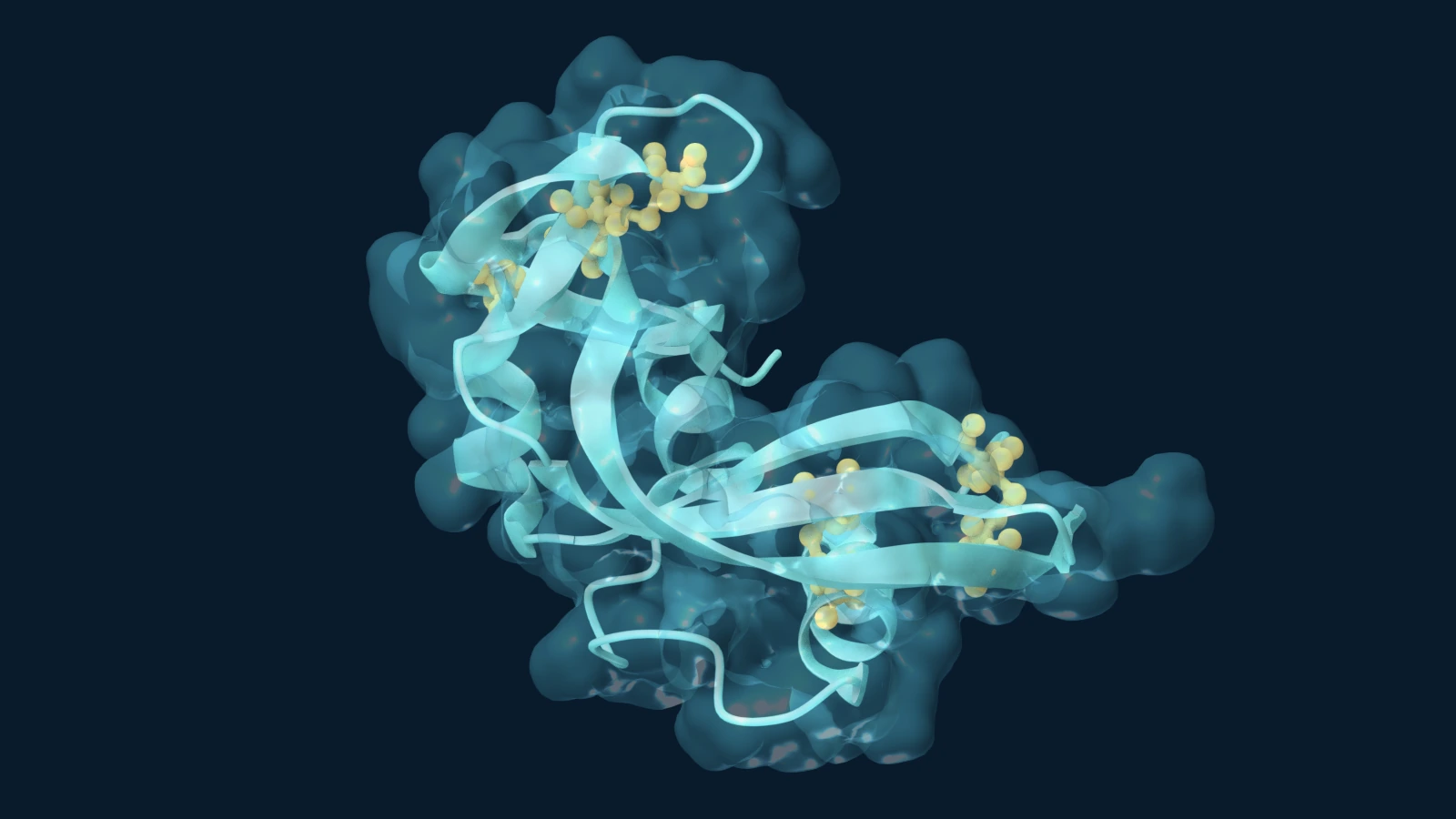
The Mol* artwork uses experimental structure PDB 7RSA: bovine RNase A determined by X-ray crystallography at 1.26 ångström resolution. Aqua ribbons trace the protein fold, while gold cysteine highlights mark the residues that form its four disulfide cross-links. A translucent molecular surface is cut away on the camera-facing side to expose the architecture beneath. Water and the crystallisation-associated tert-butyl alcohol molecule are omitted for clarity. The deposited atomic coordinates are unchanged.
The image shows the folded enzyme, not its unfolding or refolding in motion. That finished shape is the point. A routine laboratory reagent helped reveal how a sequence can specify a functional structure—and how distant parts of a chain can meet to perform chemistry. Among the enzymes in our Nobel history, few pack so much insight into so small a molecule.
Sources and further reading
- Nobel Prize: the 1972 chemistry award and its two distinct citations.
- Anfinsen, C. B. (1972). Studies on the principles that govern the folding of protein chains. Nobel lecture.
- Moore, S. and Stein, W. H. (1972). The chemical structures of pancreatic ribonuclease and deoxyribonuclease. Nobel lecture.
- RCSB PDB-101: ribonuclease A, its cutting activity and structural stability.
- NCBI Bookshelf: protein folding, chaperones and processing.
- New England Biolabs: RNase A as a reagent for removing RNA from genomic DNA preparations.
- RCSB PDB 7RSA and the primary structure study by Wlodawer and colleagues (1988).