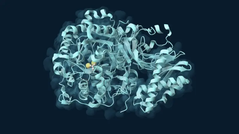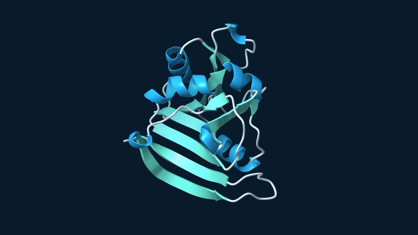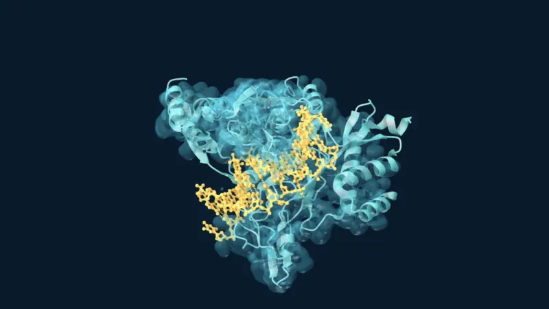
Two neighbouring thymines in DNA can survive a burst of ultraviolet light with their identities intact yet become almost unreadable. The problem is an unwanted chemical connection: two new bonds link the bases to one another. A polymerase trying to copy this stretch can stall. In organisms equipped with an enzyme called photolyase, there is a surprisingly economical way out. Instead of cutting out the damaged letters, the enzyme uses light to undo the bonds that should not be there.
It sounds contradictory. If light caused the damage, why should more light reverse it? Follow the energy through the two events and the contradiction disappears. In the first, DNA absorbs ultraviolet radiation and a particular pair of bases reacts. In the second, blue light supplies energy to a specialised molecule inside a protein that has already recognised the damage. There is no general rule that illuminating DNA makes it whole again.
What exactly has gone wrong?
Thymine is one of DNA’s four letters. Ordinarily, a thymine is attached to its sugar and can pair with adenine on the opposite strand. If two thymines sit side by side on one strand, ultraviolet exposure can link their rings, forming a four-membered ring between them. That is one example of a cyclobutane pyrimidine dimer, or CPD. “Dimer” here means that two nearby bases are joined. It does not mean that two DNA strands have been glued together.
The backbone carrying the sequence has not been cut. The two letters are still there; it is their local chemistry and geometry that have changed. CPD photolyase recognises this kind of lesion. Other kinds of ultraviolet damage exist, and the enzyme in this story is not a universal DNA repair kit. Unlike the copying reaction discussed in how reverse transcriptase copies a template, the repair needs neither a fresh stretch of sequence nor a supply of replacement nucleotides.
Why pull the bases out of the helix?
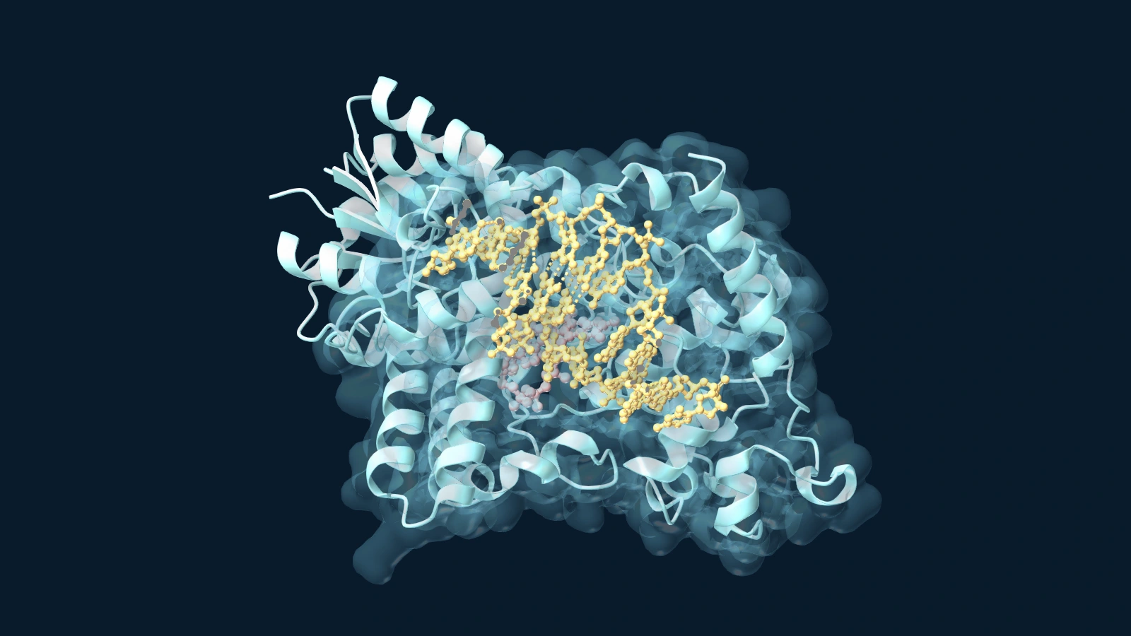
In 2004 Alexandra Mees and colleagues crystallised a cyanobacterial photolyase with a short synthetic duplex containing a CPD-like lesion. In the photolyase–DNA structure, the duplex bends and the affected pair sits outside the usual stack of bases. The enzyme has brought the pair into a pocket near its flavin cofactor, a non-protein molecule that participates directly in the chemistry. Keeping the DNA intact while turning the damage towards the cofactor solves a practical problem: the enzyme can reach precisely the bonds it needs to undo.
The image comes with a detail worth reading before interpreting it. In the original crystal experiment, exposure to synchrotron radiation at 100 kelvin split the lesion in the crystal. The authors saw separated thymines still occupying the pocket. The structure is therefore especially informative about how the enzyme grips the DNA and where the bases can sit just after repair. It would be misleading to illustrate those same deposited coordinates as an intact dimer waiting for its first blue photon.
An electron goes out and comes back
In the working enzyme, the catalytic flavin is in a reduced form called FADH⁻. Absorbing light energy raises it to an excited state from which it can transfer an electron to the damaged bases. The extra electron changes which arrangements of bonds are favourable. The two cross-links can break, leaving separate thymines. An electron then returns to the flavin. The energy input came from light; the enzyme does not have to spend a fresh flavin molecule on each repair.
That order can be tested without seeing individual atoms. In the ultrafast spectroscopy experiments, researchers excited the enzyme–substrate complex with short light pulses and followed changes in absorption at different wavelengths. Transient signals appear while the electron is away and disappear as the cofactor recovers. Such measurements can distinguish events that would all be over before a conventional reaction assay took its first reading.
Speed alone does not guarantee success. If an electron goes back before repair finishes, the flavin can recover without the lesion being repaired. The useful question is which competing events usually win. In the efficiency study, changing the protein environment and comparing reaction pathways helped explain why the timing of the whole cycle, rather than the speed of electron delivery alone, matters to the fraction of successful repairs.
Later, the 18-snapshot experiment used time-resolved crystallography to examine a CPD photolyase after light activation. Researchers identified successive changes in the lesion and the environment around it. These snapshots sample populations of crystals at different delays, not one enzyme tracked frame by frame. They supply something complementary to spectroscopy: a way to relate the timing of chemical events to positions within the pocket.
Why human cells take a different route
This is a striking piece of biology precisely because it is not universal. In the comparison of cell extracts, researchers readily detected photolyase activity in bacterial, yeast and rattlesnake samples but not in the human white blood cell or HeLa extracts tested. Human cells repair bulky UV lesions by other means, notably nucleotide excision repair.
The difference is visible in the products. In human cell experiments, investigators collected and sequenced short pieces of DNA released during excision, producing the map of excised DNA. An excision system cuts out a damaged stretch and the gap is subsequently filled. A CPD photolyase leaves the original letters in place. EnzymeNews’s guide to Nobel-recognised DNA repair puts excision alongside other ways cells maintain their genomes.
A repaired sequence does not keep a record of how it was restored. A strip of excised DNA identifies one route. Two separated bases still held next to a flavin identify another. Knowing which evidence to look for is what turns the attractive idea of “repair by light” into a mechanism that can actually be tested.